Bacterial RNA promotes proteostasis through inter-tissue communication in C. elegans
What an animal eats can quietly reshape how its cells handle protein damage — but the molecular cues that travel from a meal into distant tissues have been hard to pin down. In C. elegans, two routine laboratory bacterial diets (E. coli OP50 and HT115) produce strikingly different outcomes in worms carrying a toxic polyglutamine protein, and nobody had cleanly explained why.
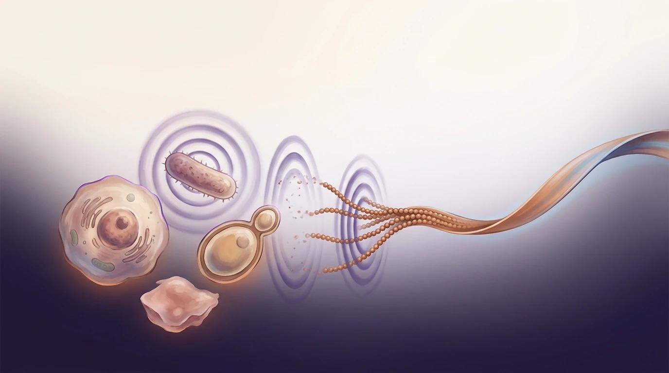
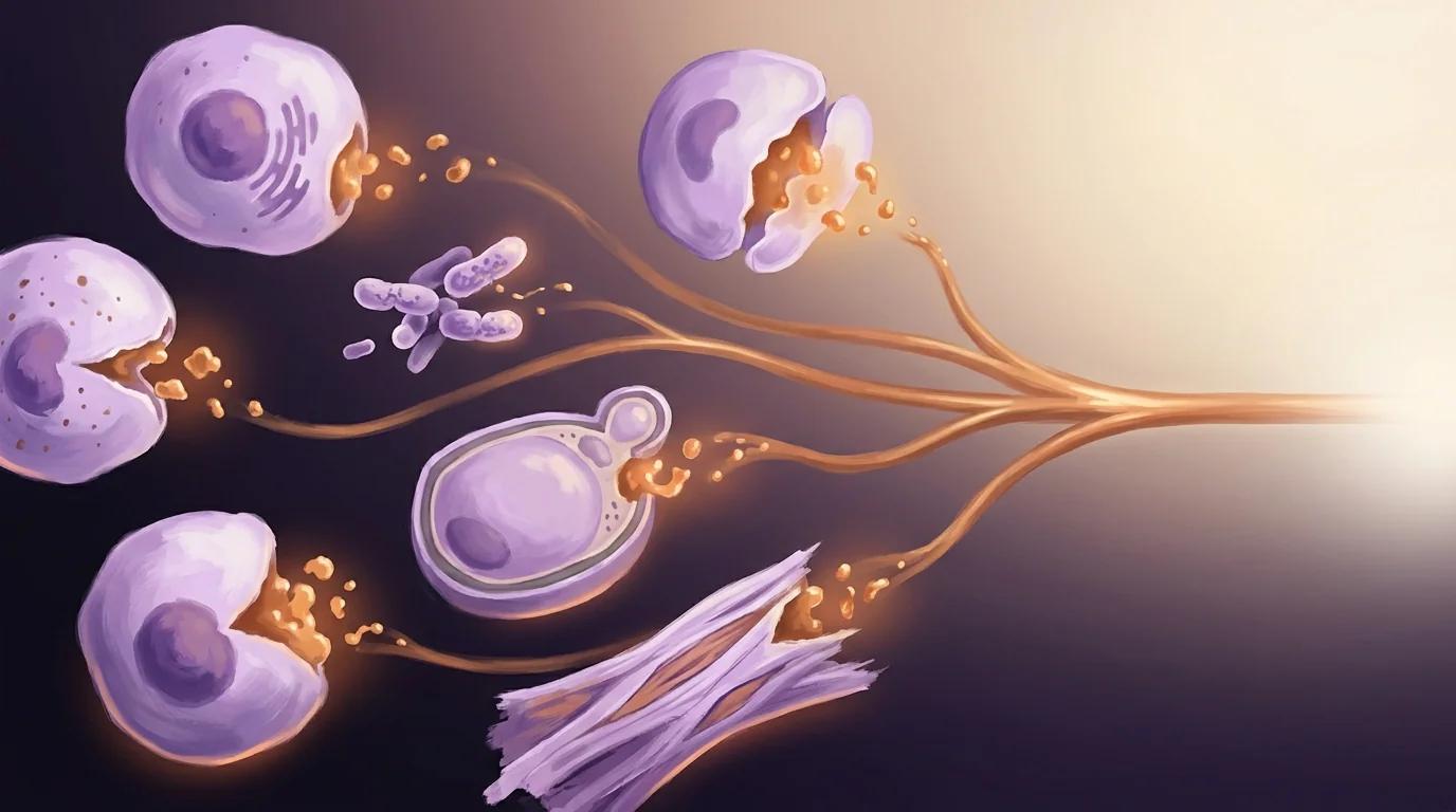
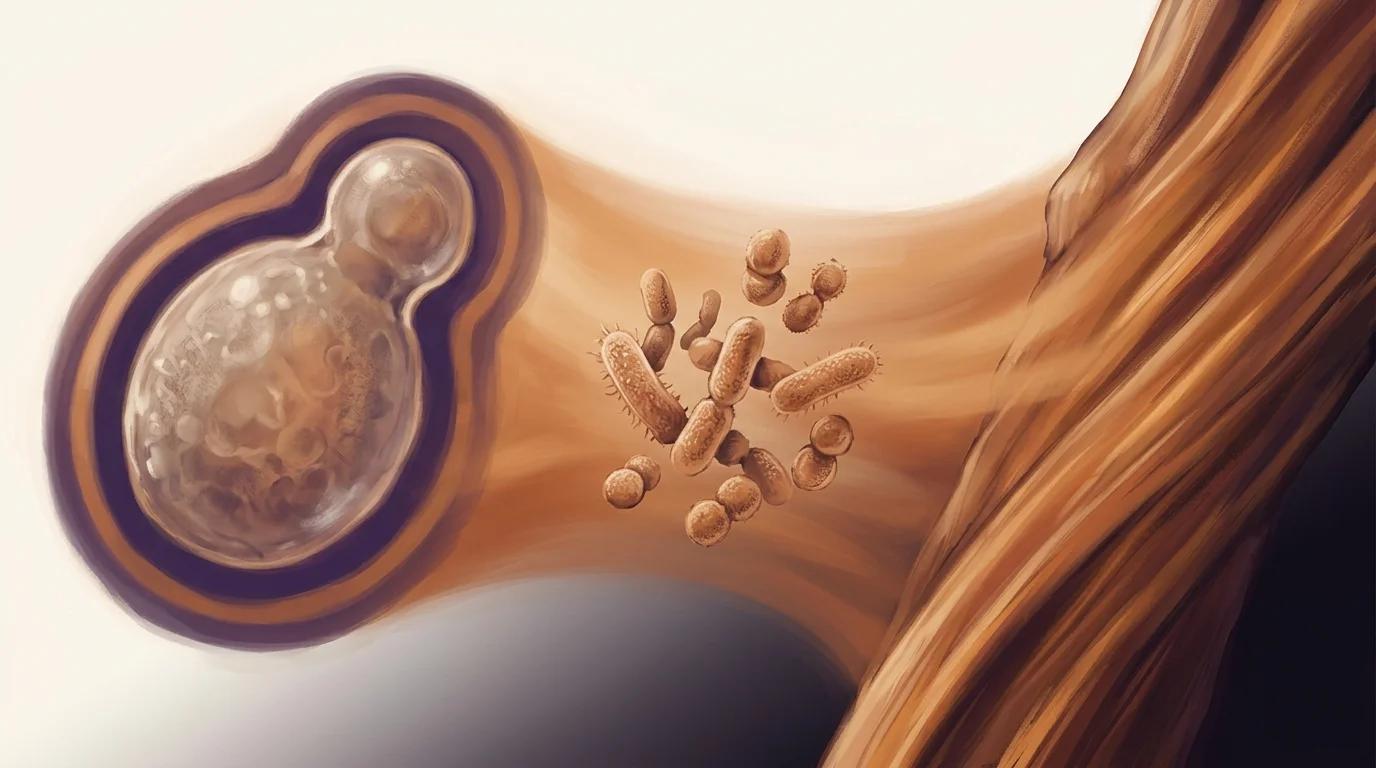
Kyriakakis and colleagues ran whole-worm DIA proteomics on worms fed three different E. coli diets and traced the protective signal back to bacterial double-stranded RNA itself. When bacteria lack ribonuclease 3, their dsRNA survives, is taken up by the worm intestine, propagates body-wide through the RNAi machinery, and triggers selective autophagy that clears protein aggregates in distant muscle cells.
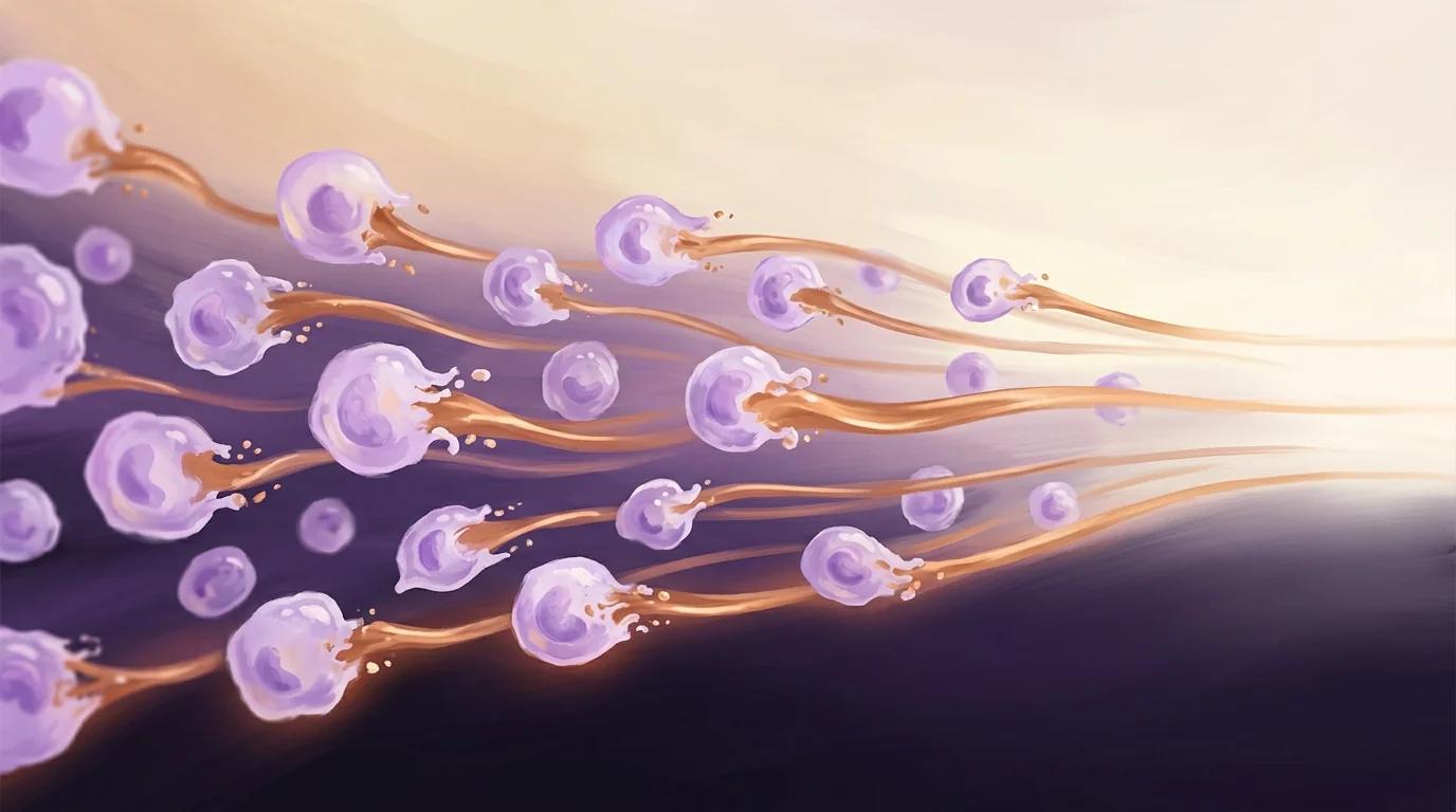
The takeaway: a dietary RNA cue flows from gut microbes to distant muscle and tunes how a whole organism handles damaged protein.
Key findings
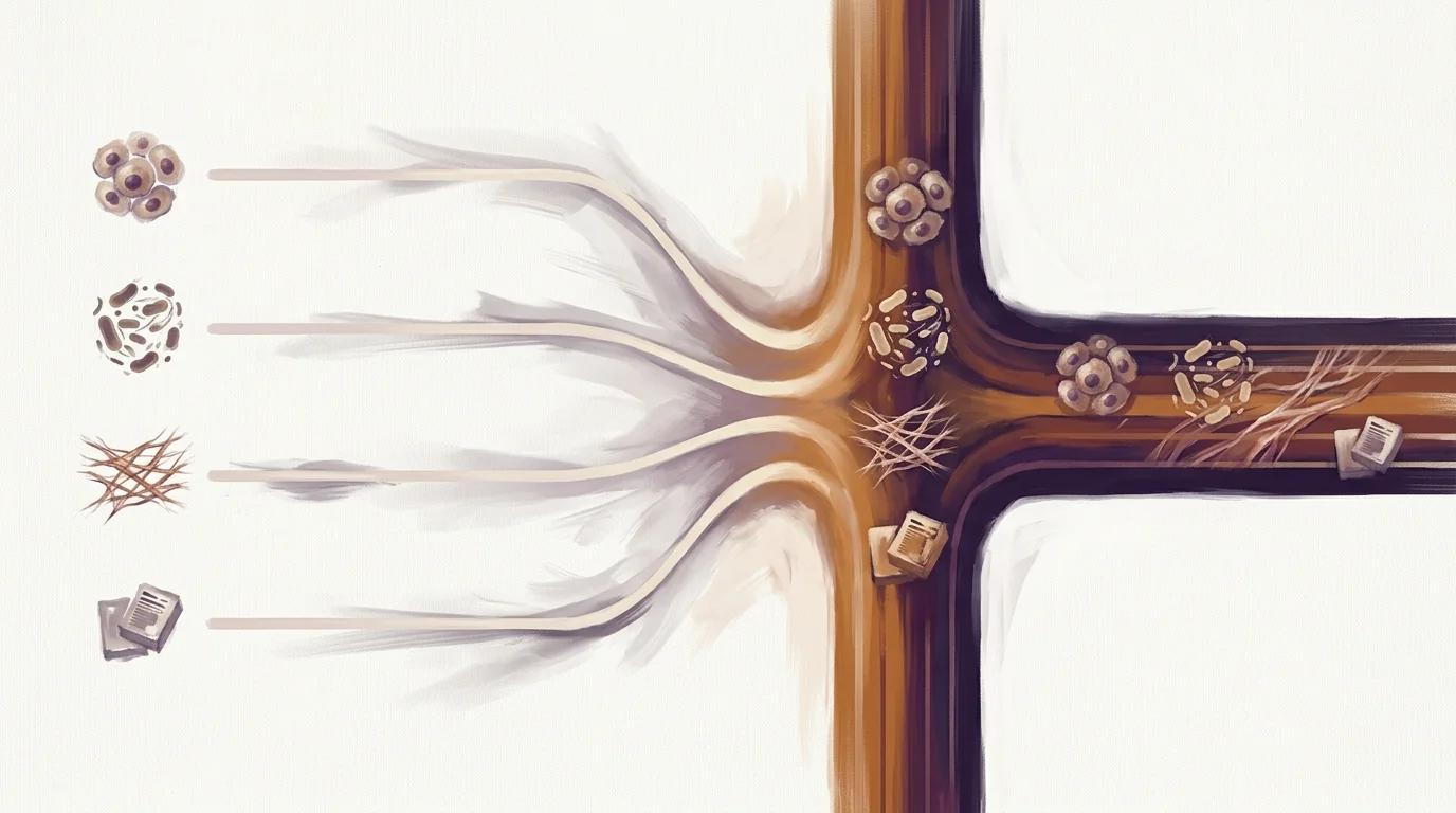
- Whole-worm DIA-LC-MS/MS proteomics on the Orbitrap Exploris 480 resolved 194 significantly altered proteins in polyQ40-expressing C. elegans fed ribonuclease-3-deficient diets versus the parental OP50 control — with a 12-protein muscle-function cluster (UNC-89, UNC-22, TTN-1, UNC-15, ATN-1, UNC-54, UNC-87, and others) that surfaces only under proteotoxic challenge, not in wild-type animals.
- Loss of a single bacterial gene — ribonuclease 3 (rnC) — protects worms from polyglutamine muscle aggregates across three independent E. coli backgrounds (HT115, OP50(xu363), and rnC-deleted Nissle 1917), with re-introduction of wild-type rnC (but not a catalytically inactive mutant) restoring aggregation — isolating dsRNA stability as the causal variable.
- The protective signal traverses three tissues: intestinal dsRNA uptake (SID-2), systemic distribution (SID-1), germline RNAi processing (PPW-1, RDE-1, ERGO-1, EGO-1), and downstream aggrephagy in body-wall muscle — knockdown of selective-autophagy receptors SQST-1, TLI-1, or WDFY-3 restores polyQ40 aggregation on the otherwise protective diet.
PIXUL in the methods
"Worms were resuspended in 5% SDS, 10 mM Tris(2-carboxyethyl)phosphine hydrochloride (TCEP), 0.1 M TEAB and lysed by sonication using a PIXUL multi-sample sonicator (Active Motif) with pulse set to 50, PRF to 1, process time to 20 min and burst rate to 20 Hz, followed by a 10 min incubation at 95 °C."
— Kyriakakis et al., Nature Communications (2025), Methods, Proteome analysis
Why it matters for PIXUL users
If you run whole-organism proteomics or study microbiome-host interactions in small model organisms, this paper is direct peer-reviewed evidence that PIXUL delivers the reproducible parallel lysis you need to resolve subtle inter-tissue biology. The authors lysed flash-frozen C. elegans pellets directly in 5% SDS / TCEP / TEAB on PIXUL, then carried the lysates straight into TCA precipitation, trypsin digestion, and DIA-LC-MS/MS on an Orbitrap Exploris 480 — one workflow, no detergent-incompatible bottlenecks, no per-sample probe cleaning, no carry-over across the three diet conditions and two genotypes compared. That consistency is what made a context-dependent 12-protein muscle cluster detectable in proteotoxically challenged animals but invisible in wild-type controls. For multi-application labs studying host-microbe biology, the practical takeaway is that the same instrument supporting your whole-organism proteomics also supports the genomics and integrated multi-omic experiments you run on the same biospecimen, while the parallel standard-microplate format matches the throughput of modern mass spectrometers and lets you scale dietary, genetic, or compound screens without re-validating sample preparation.