Comparison of the Proteome of Huh7 Cells Transfected with Hepatitis B Virus Subgenotype A1, with or without G1862T
Hepatitis B virus subgenotype A1 dominates sub-Saharan Africa and carries a sharply elevated risk of liver cancer at a younger age than other genotypes. One of its signature precore mutations — G1862T — is enriched in hepatocellular carcinoma tissue and is known to stall the HBeAg precursor in the endoplasmic reticulum, but how that bottleneck actually reshapes the host hepatocyte has been unclear.
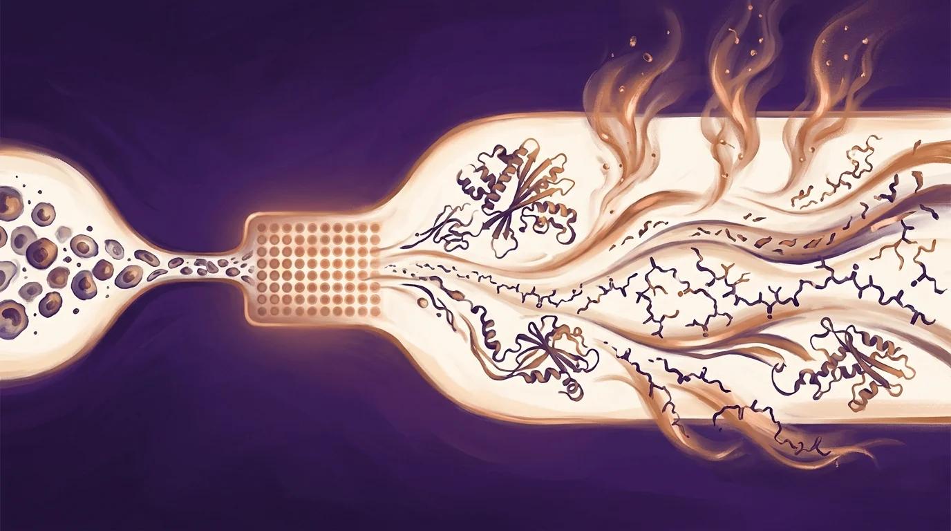
The authors transfected Huh7 liver cells with either wildtype A1 or A1 carrying G1862T and quantified the full cellular proteome against an empty-vector control by DIA SWATH-MS. G1862T uniquely dysregulated oxidative stress, cell-cycle, and MAPK signalling pathways and pushed two DNA-replication proteins (PRIM2 and RPA) above 2-fold over wildtype — a plausible molecular bridge between the mutation, its ER stress upstream, and the elevated hepatocarcinogenic risk downstream.
Key findings
- 3,906 proteins identified at 1% FDR from Huh7 cell lysates by DIA SWATH-MS five days post-transfection — a full hepatocyte-line proteome generated from PIXUL-prepared input across four biological replicates per condition.
- 247 differentially expressed proteins in wildtype A1 versus vector and 123 DEPs in G1862T versus vector, with 15 dysregulated pathways unique to G1862T spanning MAPK signalling, DNA synthesis and methylation, and extracellular matrix organisation — quantifying how the precore mutation rewires the host proteome relative to wildtype HBV.
- Oxidative stress and cell-cycle pathways were uniquely dysregulated in G1862T-transfected cells, with DNA primase subunit 2 (PRIM2) and Replication Protein A (RPA) upregulated more than 2-fold versus wildtype and confirmed by RT-qPCR plus confocal microscopy showing nuclear RPA localisation — linking the mutation to two distinct hepatocarcinogenic mechanisms.
PIXUL in the methods
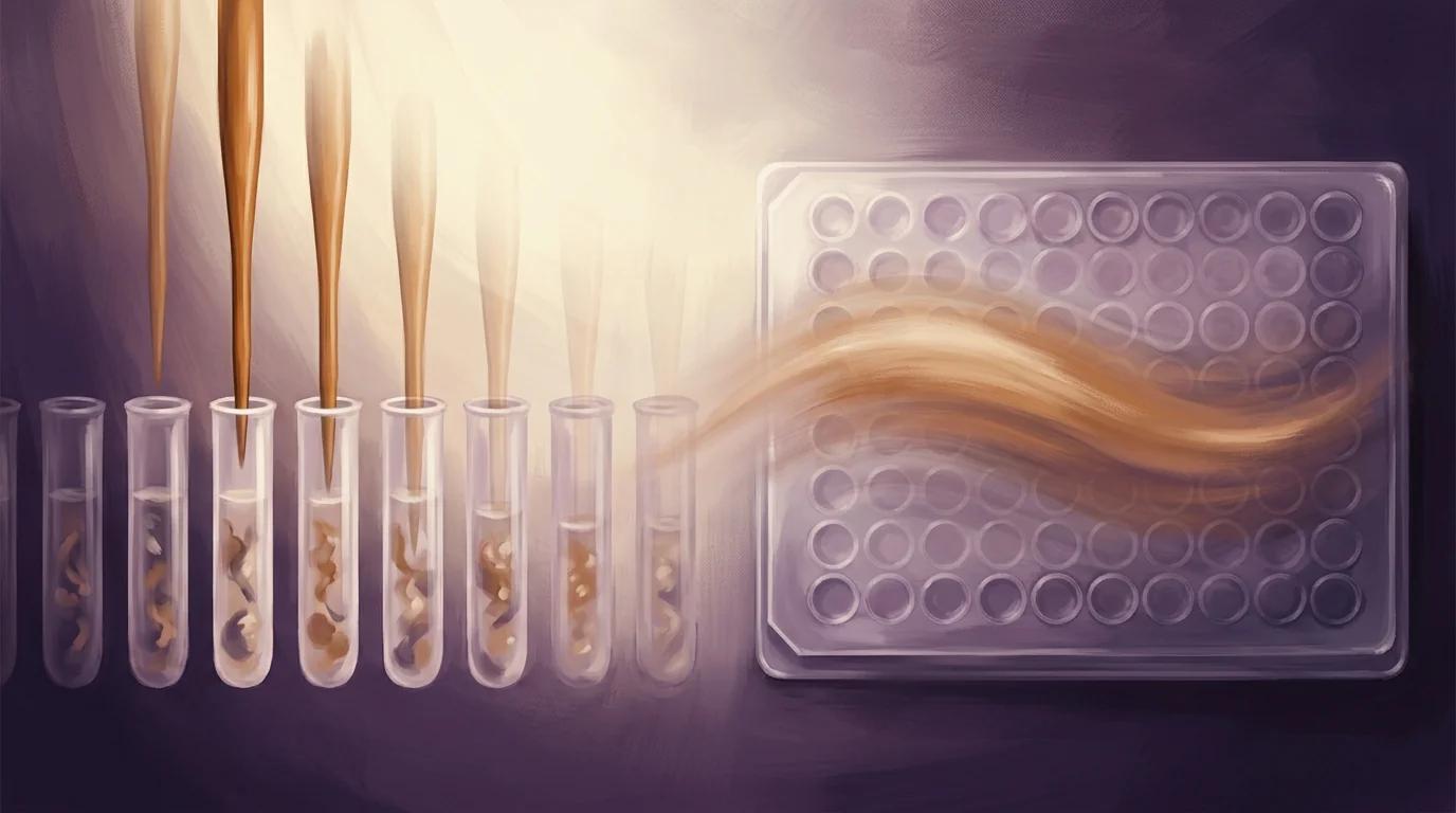
"Cell pellets were thawed on ice and resuspended in 200 μL of lysis buffer (1% SDS, 50 mM Tris-HCl pH 8.0). Protein was extracted from cell pellets using a PIXUL multi-sample sonicator (Active Motif, Carlsbad, CA, USA). Sonication settings were: Pulse = 50; PRF = 1; Process Time = 30; Burst = 20."
— Padarath et al., Current Issues in Molecular Biology (2024), Section 2.3.1, Cell Lysis and Protein Preparation
Why it matters for PIXUL users
If you run cell-line proteomics — viral transfection panels, drug perturbations, or any comparative design where biological replicates have to land on a mass spectrometer together — this paper is a direct, peer-reviewed example of PIXUL feeding a quantitative DIA SWATH-MS workflow that identifies nearly four thousand proteins per condition. The authors lyse Huh7 cell pellets in 200 μL of 1% SDS buffer, sonicate on PIXUL with a single fixed parameter set, and move directly into automated HILIC clean-up and on-bead trypsin digest. For your own viral, drug, or perturbation studies, the practical takeaway is that PIXUL delivers reproducible, parallel protein extraction at the four-replicate-per-condition statistical density these comparative designs require, without artisanal probe-tip handling between samples. One instrument, one parameter file, and an SDS-compatible lysate ready for MS-grade digestion — consistency at scale for the proteomics experiments your biology actually needs.