Comparative Proteomic Analysis of Huh7 Cells Transfected with Sub-Saharan African Hepatitis B Virus (Sub)genotypes Reveals Potential Oncogenic Factors
Hepatitis B virus is not one virus — it is a family of (sub)genotypes that circulate in different parts of the world and carry strikingly different risks of liver cancer. Sub-Saharan Africa has the highest burden, and subgenotype A1 in particular is linked to elevated hepatocellular carcinoma risk, yet the molecular reasons why have stayed elusive.
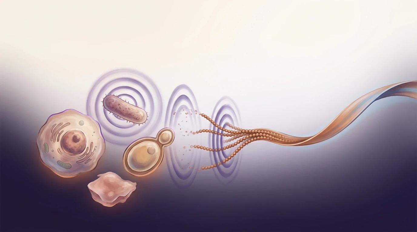
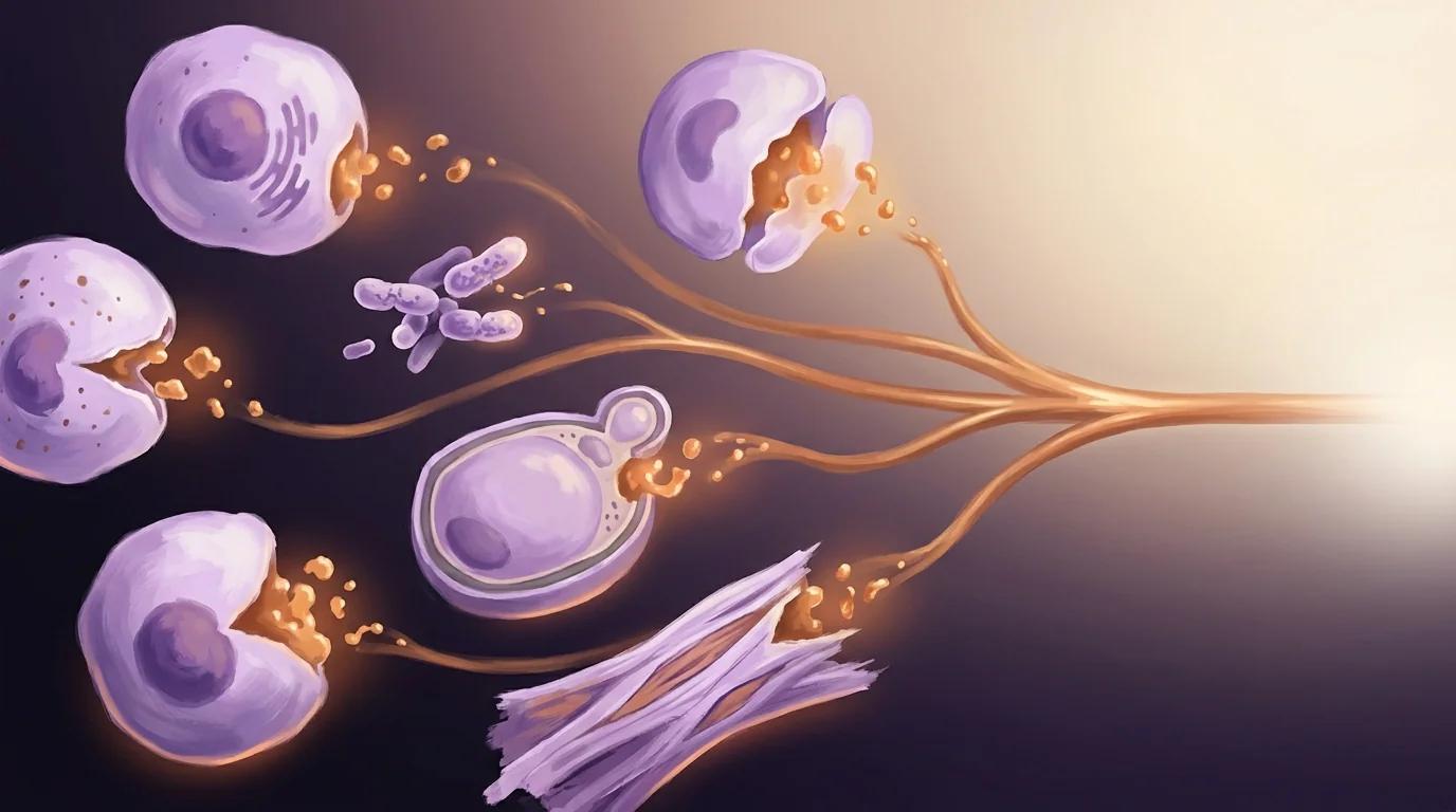
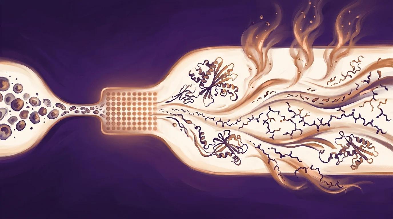
Padarath and colleagues ran a side-by-side proteomic comparison of Huh7 liver cells transfected with four HBV variants — subgenotypes A1, A2, D3, and genotype E — using PIXUL multi-sample sonication for parallel cell lysis followed by SWATH-MS quantification. Across 3,906 proteins, each (sub)genotype produced a distinct signature, and A1 uniquely upregulated three Ras-pathway regulators (RHOC, Rap2B, GNB1) tied to known oncogenic signalling.
The takeaway: HBV-driven liver cancer is (sub)genotype-specific, and parallel sample prep makes head-to-head proteomic comparisons of closely related viral strains achievable in a single experiment.
Key findings
- 3,906 proteins and 36,107 peptides quantified by SWATH-MS across Huh7 cells transfected with HBV (sub)genotypes A1, A2, D3, and E — a proteome-wide comparison of sub-Saharan African HBV variants in a human hepatocellular background, with each (sub)genotype producing a distinct expression signature relative to mock-transfected controls.
- PI3K/Akt/mTOR signalling emerged as the top exclusive oncogenic pathway in subgenotype A1 by GSEA, alongside MAPK and p53 pathway dysregulation, while Wnt/β-catenin was downregulated in A1 unlike the other variants — pointing to (sub)genotype-specific routes to hepatocarcinogenesis.
- RHOC, Rap2B, and GNB1 were upregulated >1.5-fold uniquely in subgenotype A1, with findings cross-validated by RT-qPCR and ELISA, and RHOC co-localising with HBV core protein at the cell periphery in 35% of A1-transfected cells versus 15–20% for the other variants — nominating these Ras-pathway regulators as candidate effectors of A1's elevated cancer risk.
PIXUL in the methods
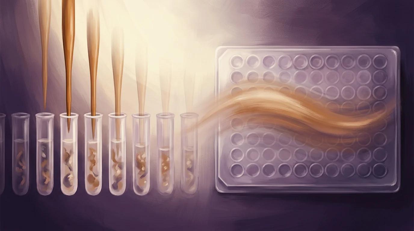
"For protein preparation, the cell pellets were thawed on ice and resuspended in 200 µL of lysis buffer (1% SDS, 50 mM Tris-HCl pH 8.0) per pellet. Protein was extracted from the cell pellets using a PIXUL multi-sample sonicator (Active Motif, Carlsbad, CA, USA). The sonication settings were as follows: Pulse = 50; PRF = 1; Process Time = 30; and Burst = 20."
— Padarath et al., Viruses (2024), Section 2.3.1, Cell Lysis and Protein Preparation
Why it matters for PIXUL users
If your work involves comparative proteomics across closely related conditions — viral strain panels, transfection series, isoform variants, oncogene allele comparisons — this paper is direct peer-reviewed evidence that PIXUL delivers the parallel-sample consistency required to resolve subtle differences between near-identical inputs. The authors lysed Huh7 cell pellets across four HBV (sub)genotypes plus controls on a single PIXUL run, then pushed those lysates through HILIC clean-up, trypsin digestion, and SWATH-MS data-independent acquisition. The result resolved 3,906 quantified proteins with enough precision to separate four near-identical HBV variants and nominate three new candidate oncogenic effectors at a fold-change cut-off as tight as 1.5x. For your own head-to-head comparisons, the practical takeaway is that single-step parallel sonication eliminates the well-to-well drift that single-sample probe sonicators introduce — drift that quietly buries the small effect sizes where biology often hides.