mTORC2 Regulates Actin Polymerization in Auditory Cells
Inner-ear hair cells are built around delicate, actin-rich structures, and when that actin scaffold breaks down, hearing follows it. The mTORC2 kinase has been a prime suspect in controlling those structures, but the wiring in auditory cells has stayed murky without a clean knockout to test it.
Lanz and colleagues built a Rictor-knockout HEI-OC1 auditory cell line to switch mTORC2 off, then ran deep proteomics on the same samples. The knockout cells lost a key PKCα phosphorylation mark, tilted their actin balance away from polymerized filaments, and shifted the abundance of more than a thousand proteins — many of them cytoskeletal.
The takeaway: mTORC2 sits upstream of an actin-regulating program in auditory cells, and deep proteomics on small specialized-cell pellets can resolve that program cleanly.
Key findings
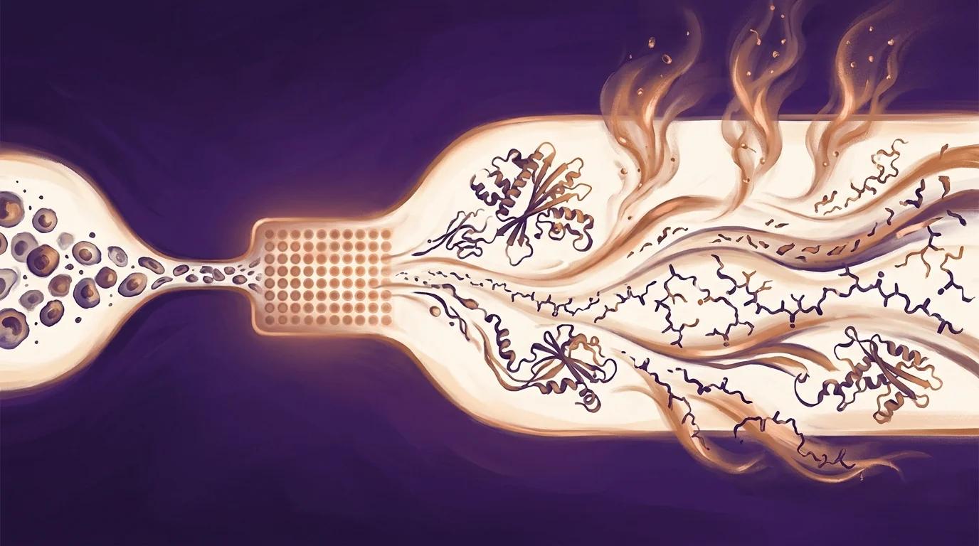
- 7,022 proteins quantified per sample across four wild-type and four RictorKO HEI-OC1 auditory cell replicates by DIA LC-MS/MS on an Orbitrap Exploris 480, resolving 611 significantly upregulated and 524 significantly downregulated proteins in mTORC2-deficient cells (log2FC > 0.6 or < −0.6, −log10 q > 2).
- Cytoskeleton, cell junctions, and extracellular matrix dominated the deregulated GO cellular components, with actin-binding and kinase activity topping molecular-function hits — direct proteomic evidence that mTORC2 loss reshapes the auditory-cell cytoskeletal proteome.
- F-actin / G-actin ratio dropped significantly in RictorKO cells (p ≤ 0.01, n = 3) alongside complete loss of PKCα Thr638/641 phosphorylation (p ≤ 0.0001), mechanistically linking mTORC2 → PKCα → actin polymerization in an auditory hair-cell model.
- Cofilin 1 (CFL1) and WDR1 — both required for stereocilia actin dynamics — were among the most strongly downregulated cytoskeletal proteins, with CFL1 downregulation independently confirmed by western blot (p ≤ 0.0001, n = 3).
PIXUL in the methods
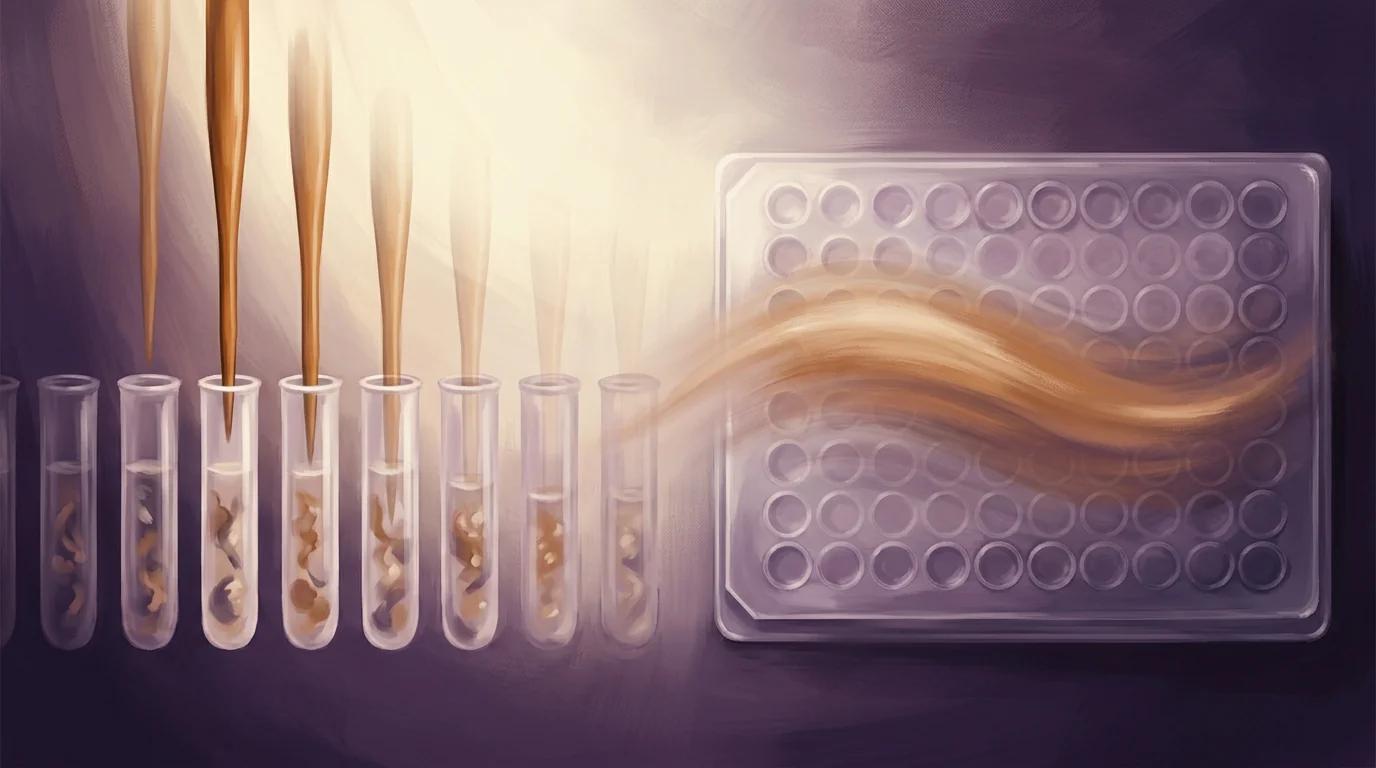
"Cells were lysed with buffer containing 100 mM TEAB pH = 8.5/5% SDS/10 mM TCEP by heating for 10 min at 95°C followed by 10 min sonication (30 s on, 30 s off per cycle) on a PIXUL system (Active Motif, CA, USA) using the following settings: pulse [N]: 50; PRF [kHz]: 1; burst rate [Hz]: 20."
— Lanz et al., Journal of Neurochemistry (2025), Materials and Methods, Section 2.11 Proteomics
Why it matters for PIXUL users
If your proteomics workflow runs on small, precious cell pellets — auditory cells, primary neurons, sorted populations, patient-derived lines — this paper is direct peer-reviewed evidence that PIXUL delivers the lysis consistency required for deep DIA proteomics on the Orbitrap Exploris 480. Lanz and colleagues quantified 7,022 proteins per sample across paired wild-type and Rictor-knockout HEI-OC1 cells with reproducibility tight enough to resolve 1,135 significantly deregulated proteins and a clean cytoskeleton-dominated GO signature. The PIXUL step ran in a defined, parameterized window — pulse 50, PRF 1 kHz, burst 20 Hz — meaning the same lysis recipe is portable across your replicates, your collaborators, and your archived experiments. For specialized-cell-type proteomic studies, the practical takeaway is that one bench-top sonicator processes up to 96 samples in parallel with non-proprietary consumables, matching the throughput your downstream mass spectrometer already expects.