Multi-omic assessment of mRNA translation dynamics in liver cancer cell lines
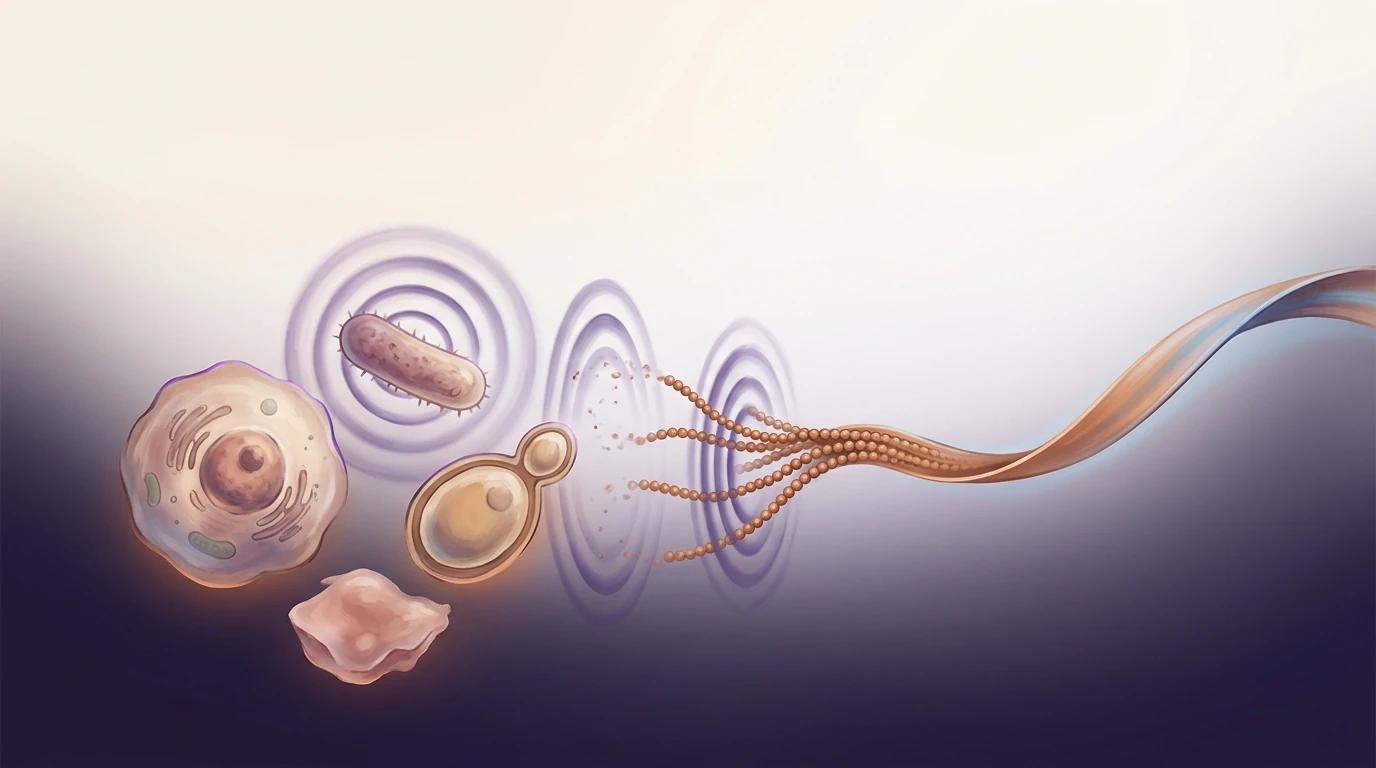
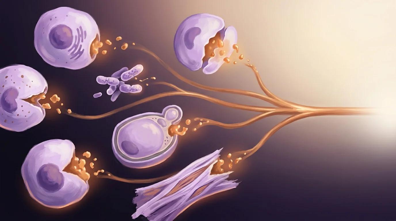
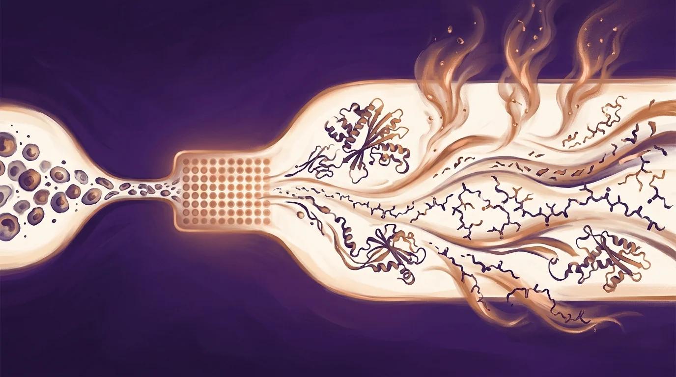
How much protein a cell actually makes from a given gene depends on a chain of steps — mRNA levels, ribosome loading, elongation rate, and final protein synthesis — that are rarely measured together in the same cells. That gap limits how well we can predict, and engineer, protein output from sequence.
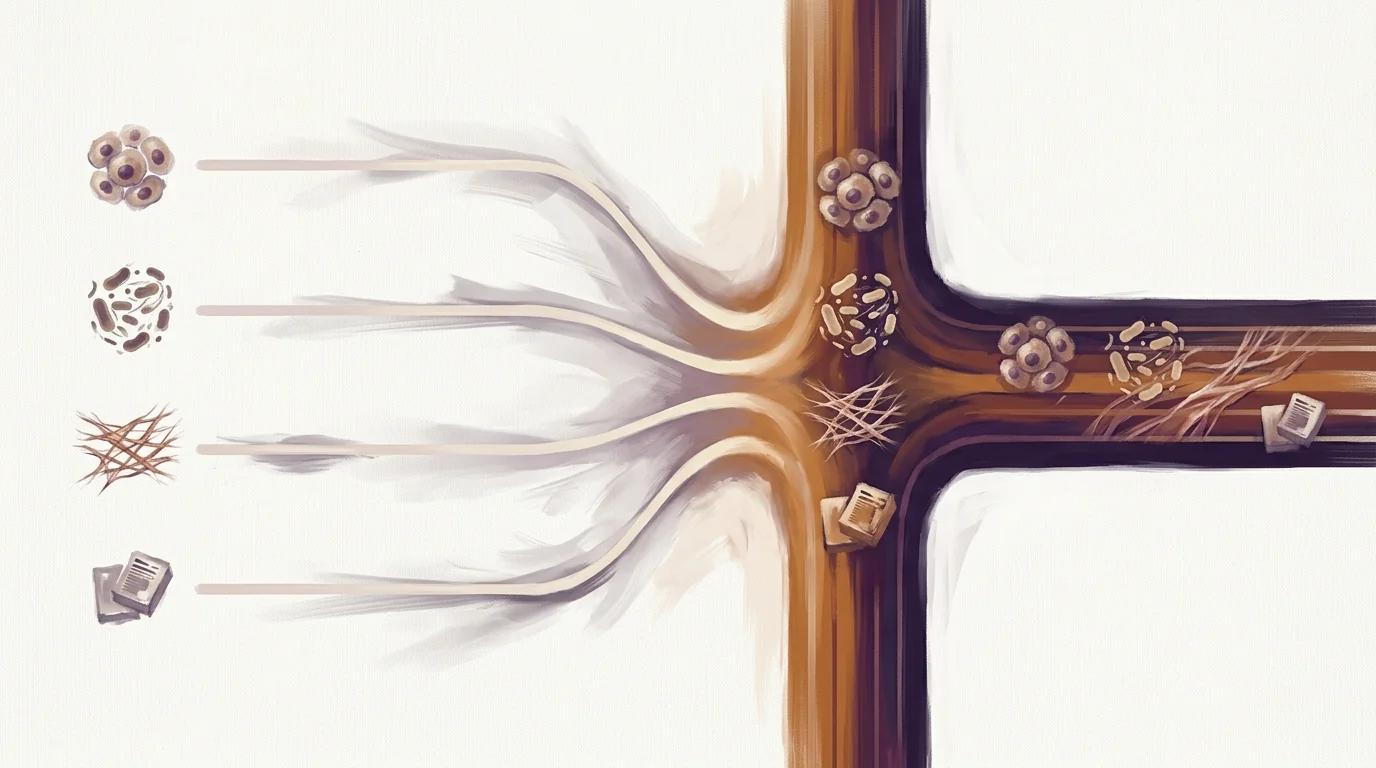
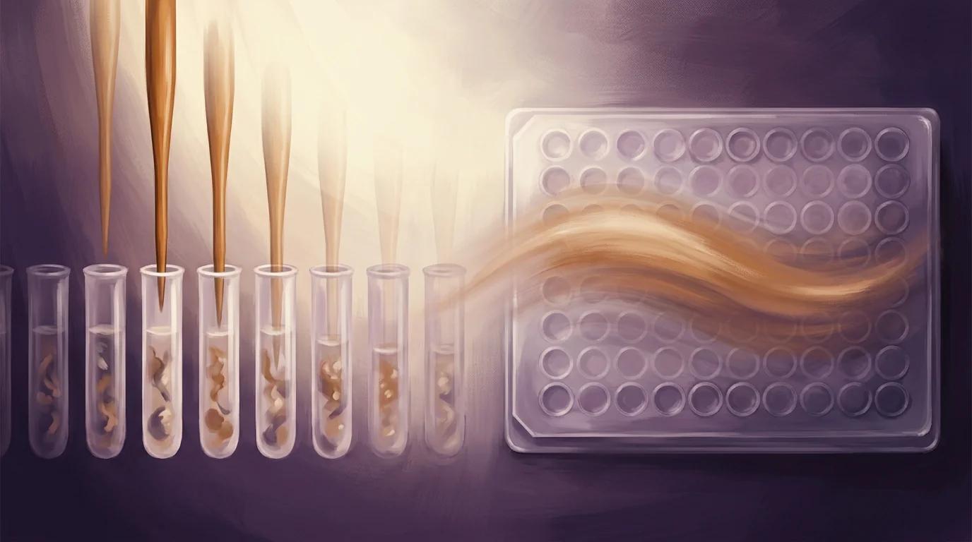
Working in two human liver cancer cell lines, HepG2 and Huh7, this team paired ribosome profiling, run-off ribo-seq, polysome profiling, RNA-seq, and pSILAC proteomics on the same samples — under both fed and nutrient-starved conditions. PIXUL handled the cell-lysis step of the pSILAC proteomics arm, keeping sample preparation consistent across the multi-omic workflow.
The result is a public multi-omic resource for liver cancer translation dynamics that the field can use to build better mRNA-to-protein prediction models.
Key findings
- Five paired translation readouts on the same HepG2 and Huh7 cells — ribosome profiling, run-off ribo-seq (harringtonine 80–180 s with cycloheximide chase), pSILAC proteomics, RNA-seq, and sucrose-gradient polysome profiling — under both standard growth and HBSS-starvation conditions.
- Average elongation rates of 4.55 aa/s in HepG2 and 4.85 aa/s in Huh7, resolved by run-off ribo-seq and consistent with prior mammalian measurements; ribosome-profiling signal explained ~50% of variance in protein abundance versus ~45% from mRNA alone.
- 89 of 92 TOP-motif mRNAs were strongly translation-suppressed under nutrient starvation, recovering the canonical translational stress response and confirming the dataset's sensitivity to physiological perturbations.
PIXUL in the methods
"Cells were lysed in 100 µl lysis buffer (5% SDS, 100 mM TEAB pH 8.5, 10 mM TCEP and 100 µg/ml CHX) using strong sonication for 10 min at 15 °C in a PIXUL sample sonicator (Active Motif)."
— Bryanton et al., Scientific Data (2025), Methods, “pSILAC proteomics sample preparation and analysis”
Why it matters for PIXUL users
If you run proteomics or multi-omic studies on cultured cells, this paper is direct peer-reviewed evidence that PIXUL fits cleanly into a stringent quantitative-translation workflow. The authors lyse HepG2 and Huh7 cell pellets in a denaturing buffer (5% SDS, 100 mM TEAB, 10 mM TCEP) with cycloheximide held at 100 µg/ml — sonication on PIXUL is the step that releases proteins from the pellet without disrupting that chemistry — and then move directly into SP3 digestion and LC-MS/MS on an Orbitrap Fusion Lumos to quantify 3,589 proteins by pSILAC. For multi-application labs working on liver cancer or any other cultured-cell system, the practical takeaway is that the same PIXUL workflow you use for proteomics also pairs cleanly with the ribosome-profiling and RNA-seq arms of the same study, so one instrument carries you from initiation kinetics to protein synthesis rates without needing a different lysis platform for each omic layer.