Quantitative proteomics of formalin-fixed, paraffin-embedded cardiac specimens uncovers protein signatures of specialized regions and patient groups
Cardiology pathology archives hold decades of formalin-fixed, paraffin-embedded (FFPE) heart biopsies — a rich record of patient disease that has historically been accessed only by histology, not by modern protein-level analysis. Formalin crosslinking and paraffin contamination have made it difficult to recover clean, quantitative proteomes from archived cardiac tissue, and prior FFPE cardiac efforts identified only a few hundred to low-thousand proteins.
Achter and colleagues built a parallel 96-well, PIXUL-based FFPE sample-preparation workflow and benchmarked it on human heart tissue. They quantified roughly 4,000 proteins per single-shot biopsy — up to 5,697 with deep TMT fractionation — and showed that formalin fixation accounted for only 1.1% of proteome-wide variance, even on blocks archived for up to fifteen years.
The takeaway: the cardiac FFPE blocks in your pathology archive are now a viable input for high-throughput, clinical-grade proteomics.
Key findings
- ~4,000 proteins quantified per single-shot FFPE cardiac sample via library-free dia-PASEF on a timsTOF HT, scaling to 5,697 proteins with >99% data completeness using deep TMT fractionation across paired FFPE and fresh-frozen left-ventricular tissue.
- Formalin fixation contributed only 1.1% of proteome-wide variance versus 28.9% for workflow choice — and matched FFPE-to-frozen samples processed with the same workflow reached Pearson R = 0.99. Archived blocks behave as quantitatively equivalent inputs.
- FFPE blocks archived up to 15 years retained proteome integrity, with no significant relationship between storage time and number of proteins identified (R = 0.093) across a 48-biopsy donor cohort.
- A retrospective cohort of 20 endomyocardial biopsies (10 arrhythmogenic cardiomyopathy patients + 10 donor controls) recovered 167 differentially abundant proteins — fibrosis collagens, TGF-β modulators, and cardiomyocyte protein losses — mirroring the histological fibro-fatty hallmark of ACM and cleanly separating disease state by PCA.
PIXUL in the methods
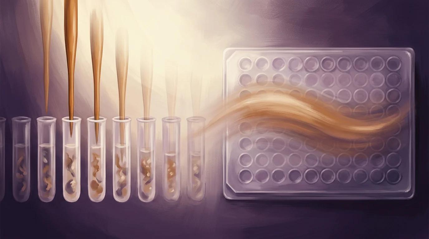
"The resulting pieces were transferred to a 96-well plate containing 100 µl of lysis buffer (5% SDS, 100 mM Tris, pH 8.5). Samples were allowed to rehydrate overnight followed by a single round of sonication using a PIXUL multi-sample sonicator for 30 min (50 N pulse, 1 kHz, 20 Hz burst rate). The plate was briefly centrifuged before being placed on a heat block for 1 h at 80 °C. After another spin-down step, sonication was repeated for an additional 30 min or until completely homogenized."
— Achter et al., Nature Cardiovascular Research (2025), Methods, Proteomic sample preparation — Protein extraction from FFPE tissue
Why it matters for PIXUL users
If your lab runs cardiology proteomics, translational research, or any retrospective study built on FFPE archives, this paper is direct peer-reviewed evidence that PIXUL anchors a parallel 96-well FFPE sample-preparation workflow capable of recovering ~4,000 proteins per single-shot cardiac biopsy — and up to 5,697 with deep TMT fractionation — from human heart blocks archived for up to fifteen years. The authors quantify exactly what you need to plan a retrospective cohort: formalin fixation contributes only 1.1% of proteome-wide variance, FFPE-to-frozen sample correlation reaches R = 0.99 under matched workflows, and block storage time shows no significant relationship with protein identification rates (R = 0.093). For your core facility or translational cardiology program, the implication is concrete: the FFPE archive sitting in your pathology department is no longer an inaccessible resource — it is now a high-throughput biobank for disease-state proteomics, regional sub-anatomical mapping, and patient-stratification studies, all running through one parallel sample-preparation instrument at a cadence that matches your downstream mass spectrometers.