SWATH-MS reveals tissue-specific proteomic changes in a Leigh syndrome mouse model
Leigh syndrome is a severe paediatric mitochondrial disorder driven by respiratory Complex I deficiency, most commonly modelled in mice by knocking out the Ndufs4 gene. The disease produces a spectrum of clinical phenotypes — ataxia, cardiomyopathy, visual problems, psychomotor regression — but the molecular response across different affected organs has not been mapped side by side.
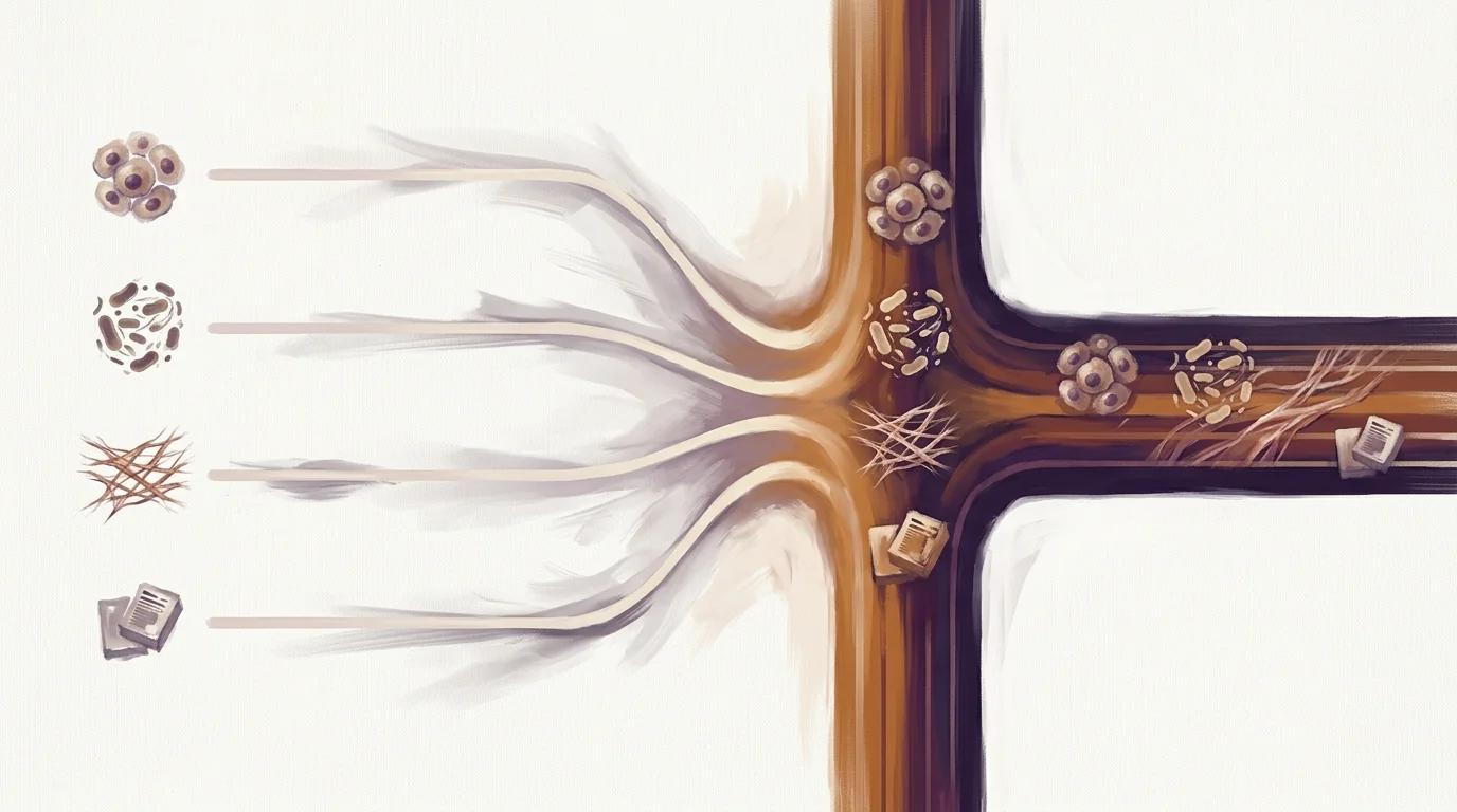
The authors used SWATH-MS quantitative proteomics to compare six tissues (brainstem, cerebellum, olfactory bulb, heart, kidney, liver) from Ndufs4 knock-out versus wild-type mice. Each organ produced a distinct proteomic signature, with the olfactory bulb and heart most disrupted, and PTEN transcriptional regulation surfaced as a candidate neurodegeneration mechanism.
Key findings
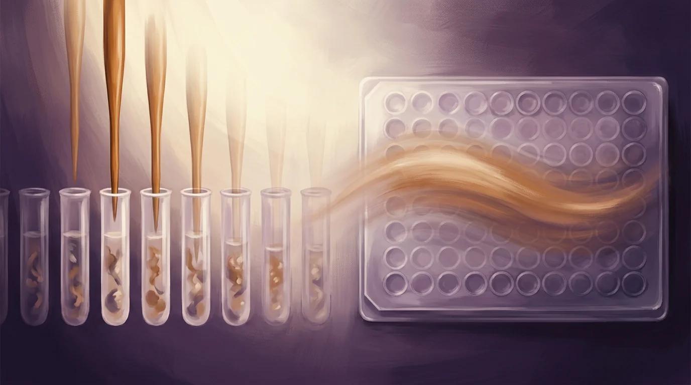
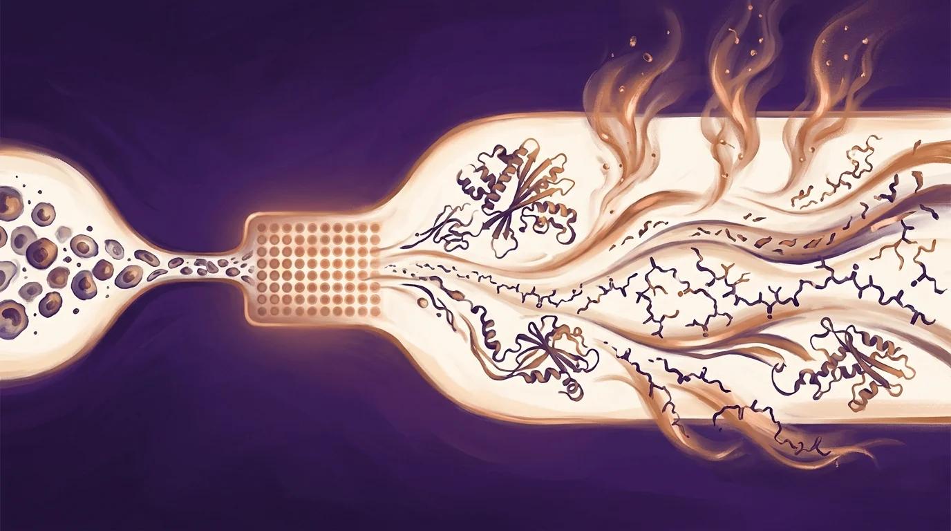
- Over 3,000 proteins quantified per tissue across six mouse organs by SWATH-MS, with all six tissue types homogenised and lysed in parallel from a single 96-well plate prior to LC-MS/MS.
- Tissue-specific differentially abundant protein (DAP) counts ranged from 75 to 189 between Ndufs4 KO and wild-type mice, with the olfactory bulb and heart showing the greatest proteomic disruption.
- Only NDUFS4 and NDUFA12 were shared as differentially abundant across all six tissues, while functional enrichment surfaced amino-acid metabolism shifts in cardiac tissue, enhanced mitochondrial translation in kidney, altered detoxification pathways in liver, and PTEN transcriptional regulation as a candidate neurodegeneration mechanism.
PIXUL in the methods
"PIXUL multi-sample sonication was used to homogenise and lyse all six tissue types in a single 96-well plate prior to SWATH-MS analysis."
— Khumalo et al., Molecular Genetics and Metabolism (2026), Methods (publisher full-text paywalled; quote from indexed abstract record)
Why it matters for PIXUL users
If your proteomics workflow has to handle multiple tissue types in the same study — rare-disease mouse cohorts, multi-organ phenotyping, DIA-MS / SWATH panels — this paper is peer-reviewed evidence that PIXUL delivers consistency at scale across heterogeneous tissues from a single 96-well plate. The authors lysed six biologically distinct mouse organs (heart, kidney, liver, brainstem, cerebellum, olfactory bulb) using one PIXUL protocol in parallel, then quantified more than 3,000 proteins per tissue by SWATH-MS with reproducibility tight enough to resolve organ-specific differentially abundant proteins down to single-tissue resolution. For translational labs running DIA-MS workflows on rare-disease tissue panels, the practical takeaway is that one instrument supports the full multi-tissue proteomic pipeline without proprietary consumables, and the parallel 96-well format matches the throughput of downstream LC-MS/MS — turning multi-organ proteomic atlases of disease models from an instrument-juggling exercise into a single-plate experiment.